
Altered brain support cells drive Dravet syndrome symptoms in mice
Study: These astrocytes could be a potential target for future treatments
Written by |


Astrocytes change how they connect and function in a mouse model of Dravet syndrome — even without visible brain damage — which likely worsens seizures and cognitive problems, making them a potential target for future treatments, according to a study.
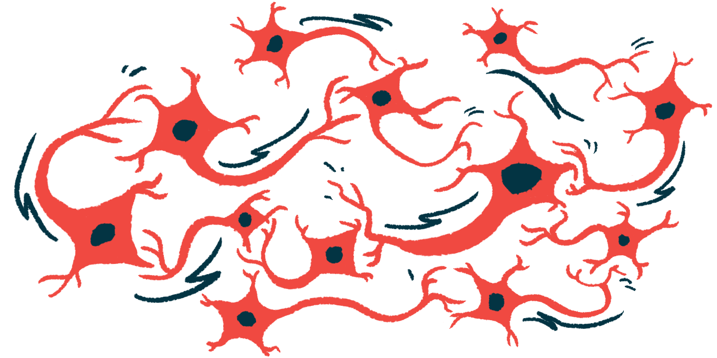
These support cells in the brain help control the environment around neurons and regulate their activity.
“Astrocytes participate in the progression of experimental [Dravet syndrome], exhibiting phenotypes [observable characteristics] that evolve over time, from early morphological alterations to long-term network-level changes,” researchers wrote in “Long-lasting remodeling of astrocytes in an Scna1+/− mouse model of Dravet syndrome,” which was published in Epilepsia.

Dravet syndrome mainly caused by mutations in SCN1A gene
Dravet syndrome is mainly caused by mutations in the SCN1A gene, which encodes a voltage-gated sodium channel, a protein that helps neurons (nerve cells) generate electrical signals. When this protein does not function properly, neurons become overly excitable, leading to frequent seizures beginning in early childhood and long-term problems with learning and behavior.
This study focused on astrocytes, which help control the chemical environment of the brain, regulate neuronal communication, and maintain balance. Their role in Dravet syndrome is still not well understood, so researchers examined how these support cells change during the course of the disease.
To do this, they used a mouse model with one mutated copy of the Scn1a gene. These mice develop symptoms similar to those of Dravet syndrome. The researchers studied different stages of the disease, from early worsening of seizures to later stabilization, using brain recordings and behavioral tests.
Model mice exhibited seizures and long-term behavioral changes as the disease progressed. Brain activity recordings confirmed severe seizures involving both the cortex and hippocampus, which are regions important for learning and memory. These seizures are often repeated within short periods.
Behavioral tests showed that, compared with healthy mice, model mice were more active, suggesting hyperactivity. They also performed worse on memory tasks, such as navigating a maze, indicating problems with working memory. Some behaviors resembled “autism-like traits,” the researchers noted, including reduced interest in typical social exploration and repetitive actions.
Despite these functional problems, the brain structure remained mostly unchanged. There was no major tissue damage, such as hippocampal sclerosis, which is scarring of brain tissue often seen in epilepsy. This means that severe symptoms can occur without visible large-scale brain damage.

Findings suggest astrocyte shape changes are temporary
The researchers then looked at astrocyte activity. They measured GFAP, a protein used as a marker of astrocyte activation. Higher GFAP levels indicate that astrocytes are reacting to stress or disease. These levels increased during the worsening phase of the disease and remained higher thereafter.
During early disease stages, astrocytes became more complex in shape, with more branches. This is called reactive gliosis, a process where support cells change in response to injury or abnormal activity. However, this increased branching did not persist in the long term, suggesting that astrocyte shape changes are temporary.
Another type of brain cell, microglia, which are involved in immune responses, showed only short-term activation. Over time, microglia returned to normal, and there was little evidence of ongoing inflammation. This suggests that chronic inflammation is not a major driver of long-term disease changes.
Astrocytes communicate through gap junctions, channels formed by proteins called connexins that allow cells to exchange ions and small molecules, helping maintain balance in the brain. In the long term, astrocytes formed larger and more connected networks. Connexin levels increased, suggesting that astrocytes reorganize as the disease progresses.
However, not all astrocyte functions improved. Hemichannels, which are half of a gap junction and allow exchange with the outside environment, worked less effectively. Impaired hemichannels may affect how astrocytes release signaling molecules and regulate neuronal activity.
Finally, the researchers studied how neurons communicate in this altered environment. They found that basic synaptic transmission was normal, but short-term increases in signal strength after stimulation, called posttetanic potentiation, were stronger. This suggests that more of the neurotransmitter glutamate was being released. Neurotransmitters are a type of chemical messenger that nerve cells use to communicate.
Overall, “astrocytes undergo long-term remodeling independent of tissue damage,” the researchers concluded. These changes include temporary shape alterations, long-term network expansion, and functional impairments. Together, these effects may contribute to seizures and cognitive problems by altering how brain cells communicate and maintain balance.
Leave a comment
Fill in the required fields to post. Your email address will not be published.