
Patient-derived Dravet models may help study disease variability
Protein patterns differed across early-stage neurospheres
Written by |


Neurospheres, 3D clusters of early-stage neural cells derived from people with Dravet syndrome, showed patient-specific protein patterns that varied in association with disease severity in a recent study.
Researchers broadly analyzed proteins in the neurospheres and found that, despite a shared molecular foundation, each patient’s cells exhibited distinct patterns of protein abundance. The cell line from the patient with the highest clinical severity score stood out as a molecular outlier, the data showed.
“These findings establish patient-derived neurospheres as a scalable human model for investigating molecular variability in [Dravet syndrome],” researchers wrote.

Patient-derived models may show molecular differences
The study, “Urine-Derived iPSC Neurospheres Uncover Proteomic Correlates of Clinical Severity in Dravet Syndrome,” was published in the Journal of Neurochemistry.
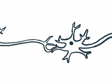
Most cases of Dravet are caused by mutations in the SCN1A gene, which impair the function of the NaV1.1 sodium channel. Because this channel is critical for inhibitory brain cells that help keep nerve signaling in balance, disruptions can lead to uncontrolled nerve cell activity, causing seizures, cognitive impairment, and motor problems.
Dravet can affect patients differently, even among those who share SCN1A mutations. Seizure frequency and severity can vary, as do cognitive outcomes, ranging from mild to profound intellectual disability. In fact, studies have yet to find a reliable link between the specific type of SCN1A mutation and overall disease severity.
Much of what’s known about the condition’s underlying molecular mechanisms has been established in animal models. Still, biological differences between mice and humans can limit the translation of findings into patient care. While human stem cell models have advanced research, flat, 2D cultures lack the complexity of real brain tissue.
Three-dimensional neurospheres grown from patient-derived stem cells may offer a more tissue-like alternative. They model early stages of brain development rather than mature nerve cell circuits, which makes them useful for Dravet research because the disease involves disruptions to early developmental processes.
Study used neurospheres from 3 patients
Scientists in Brazil explored whether neurospheres derived from three people with Dravet — each carrying a distinct SCN1A mutation — could reveal molecular differences that may help explain the wide clinical variability of Dravet.
The first patient (DRVT1) had a deletion involving two SCN1A gene segments but experienced mild disease, as indicated by the caregiver-assessed Dravet Syndrome Neurodevelopmental and Comorbidity Evaluation (DANCE) checklist. It evaluated four areas: cognition and behavior, motor abilities, daily living skills, and family quality of life.
The second (DRVT2) had a mutation that led to a shortened protein and experienced very high disease severity, with pronounced difficulties across cognition, behavior, motor function, and family quality of life. The third (DRVT3) had moderate disease and carried a mutation in SCN1A, as well as an additional mutation in SCN9A, a gene that has been proposed as a potential modifier in epilepsy and Dravet. DRVT3’s challenges were mainly related to cognition and behavior.
The team broadly analyzed proteins from patient-derived neurospheres. Across all three lines, the distribution of protein abundance was highly similar, meaning that the neurospheres shared a common molecular foundation. Yet patterns distinguishing each patient line from the others also emerged.
Analysis of overlapping differences consistently identified DRVT2, the cell line from the patient with very high disease severity, as a molecular outlier, whereas DRVT1 and DRVT3, the mild and moderate lines, showed more similar abundance profiles. The researchers noted that these comparisons describe variability within this small group and are not intended to define changes related to Dravet in general.
Protein patterns differed with disease severity
Proteins found at higher relative abundance in DRVT1 (mild) were associated with processes involved in synapses, the junctions between two nerve cells that facilitate communication. On the other hand, DRVT2 (very high severity) showed a higher relative abundance of proteins associated with RNA processing and a lower abundance of proteins associated with mitochondrial energy production.
Overall, protein patterns aligned with milder clinical severity were related to synaptic function, mitochondrial energy production, and cell adhesion (the ability of cells to stick together). Greater disease severity was associated with protein patterns related to RNA processing and protein quality control.
Researchers then mapped differentially abundant proteins onto established synaptic pathways. Compared with DRVT1, both DRVT2 (very high severity) and DRVT3 (moderate) showed lower relative abundance of proteins involved in the release of synaptic vesicles, the tiny sacs that contain neurotransmitters (nerve signaling molecules). Several of these proteins were involved in neurotransmitter release and synaptic plasticity, the ability of synaptic connections to strengthen or weaken over time.
“Our findings indicate that patient-derived neurospheres can reflect inter-patient molecular differences within [Dravet syndrome] and that proteomic profiles vary in association with clinical severity measures in this cohort,” the scientists concluded. The researchers said the model could provide a foundation for future studies linking molecular profiles to clinical variability in Dravet syndrome.


Leave a comment
Fill in the required fields to post. Your email address will not be published.