Inhibitory Cells Can’t Send Signals to Neighboring Cells in Dravet Mice

A type of brain nerve cells, called parvalbumin-expressing (PV) interneurons, was able to generate electrical signals in a mouse model of Dravet syndrome, but failed to properly transmit them to neighboring cells, according to a recent study.
This failure in a process called synaptic transmission may underlie the symptoms of Dravet in adult mice, the researchers suggested.
Moreover, the team said, its earlier emergence in some adolescent mice could contribute to their premature death, called sudden unexpected death in epilepsy (SUDEP).
“We provide evidence that [PV interneurons in Dravet mice] exhibit impaired synaptic transmission, that early impairments in [PV interneuron] synaptic transmission may predict SUDEP risk, and that such deficits persist across development and may contribute to the defining pathology” of the disease, the researchers wrote.
The study, “Developmentally regulated impairment of parvalbumin interneuron synaptic transmission in an experimental model of Dravet syndrome,” was published in Cell Reports.
Mutations in the SCN1A gene that lead to a loss of function of the sodium channel subunit Nav1.1 are the cause of Dravet syndrome. In particular, the loss of Nav1.1 in PV interneurons, which are important for seizure suppression, is thought to underlie Dravet’s hallmark symptom of severe, treatment-resistant seizures.
“Dravet syndrome affects 1 in 14,000 children in the world and has a profound impact on children and their families,” Ethan Goldberg, MD, PhD, the study’s senior author, said in a press release.
“We can model Dravet syndrome in the laboratory to understand precisely how the loss of SCN1A produces the clinical features characteristic of the disease to drive development of novel therapies, and, one day, a cure,” said Goldberg, a pediatric neurologist and director of the Epilepsy Neurogenetics Initiative (ENGIN) at the Children’s Hospital of Philadelphia.
Data from mice that lack normal levels of Nav1.1 — a model of Dravet — have shown that, early in development, PV interneurons are unable to fire electrical signals properly. Some evidence suggests, however, that this impaired firing normalizes later on, although symptoms persist.
To investigate this phenomenon, researchers at the Children’s Hospital of Philadelphia, led by Goldberg, evaluated changes in PV interneuron activity at early disease stages, and then again at later disease stages in the same mouse model.
When the mice were young (16-21 days old), the researchers took a small biopsy of brain tissue and examined the PV interneurons’ electrical properties.
They found that all Dravet mice exhibited reduced electrical firing rates of PV interneurons compared with healthy mice of the same age (a control group). These findings suggested impaired generation of electrical signals in the PV cells.
After these first recordings, 26.2% of the mice experienced SUDEP and did not survive to young adulthood (35 days old), whereas all control mice did.
PV interneuron activity was again analyzed in brain slices from the mice that did survive into young adulthood (35-56 days old). As expected, normal electrical signals were now generated in the PV interneurons, but a process called synaptic transmission appeared impaired.
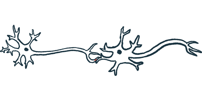
Synapses are the junctions between two nerve cells that allow them to communicate. Normally, a cell sends an electrical signal down its own axon, or projection, which is then converted to a chemical signal that is used to communicate with neighboring cells. In the case of PV interneurons, this process is used to inhibit nearby excitatory cells, whose excessive activity leads to seizures.
Excitatory and inhibitory signals are responsible for maintaining adequate stimulation of brain nerve cells. Excitatory signaling from one nerve cell to the next makes the latter cell more likely to fire an electrical signal while inhibitory signaling makes the latter cell less likely to fire.
The researchers found that PV interneurons in adult Dravet mice could generate the electrical signal. However, these cells failed to properly send the signal down their axons, thereby stalling the chemical communication needed to inhibit nearby cells.
PV interneurons in these mice also formed fewer physical connections with excitatory cells.
In younger mice, these synaptic deficits were only observed in the group of mice that died before reaching adulthood.
The team next used a technique called optogenetics to specifically stimulate PV interneurons at the soma — where an electrical signal is generated — or at the synapse, which is where a chemical signal is generated. They then measured the response of the nearby excitatory cells.
In young mice that later died, as well as surviving adult mice, responses to synaptic stimulation were markedly diminished, again suggesting synaptic transmission deficits.
The results overall suggest that impairments in synaptic transmission contribute to ongoing disease symptoms even after electrical firing of the PV interneurons is restored. The combination of electrical dysfunction and early synaptic impairment also may contribute to SUDEP in some young mice, the results also suggested.
“We conclude that combined dysfunction of PV-[interneuron] spike generation and synaptic transmission drives disease severity, while ongoing dysfunction of synaptic transmission contributes to chronic pathology,” the researchers wrote.
The findings also inform new treatment strategies, the researchers said, noting that new approaches should be aimed at restoring PV interneuron synaptic transmission. “A prediction of our work is that the success of therapies under development may depend on the ability to increase expression of Nav1.1 at the parvalbumin interneuron axon,” Goldberg said.


